Cholera Toxin


[Sources and Historical Background] [The Culprit Protein] [At the Molecular Level] [Effects] [Treatment] [References and External Links]

Sources and Historical Background

Image courtesy of NSF
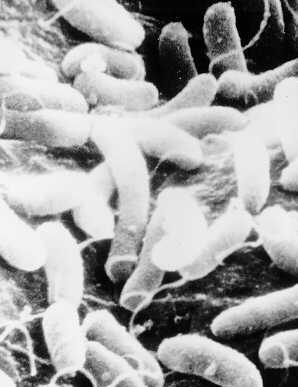
Cholera toxin is produced by the bacterium Vibrio cholerae, shown at left. It is a gram negative curved rod (hence vibrio) with a single polar flagellum. This flagellum allows the bacterium to be motile, thus it can "swim" against a current. The flagellum can also be a tool of attachment to host tissues in some cases. It is a facultative anaerobe, meaning its metabolism is adapted to function in both aerobic and anaerobic conditions. It is quite common and is often found on surface waters, both fresh and saltwater.

Proper water filtration systems and effective sewage treatment facilities ensure that V. cholerae does not cause problems with humans. It is for that reason that Cholera cases are few in the United States and most developed nations. Most cases that do occur in developed nations can often be traced back to foreign travel to underdeveloped nations with an unclean drinking water supply. However, without proper sanitation, Cholera can easily affect humans. If left untreated, it has a fatality rate of up to 50%. However easy means of treatment, explained later, lower the fatality rate to 1% in adults and 2% in children.

Epidemic cholera, as it is often called, has been affecting humans at epidemic levels for centuries. One particularly interesting case is the Broad Street Pump Outbreak in London, England. In the summer of 1854, cholera epidemics had been sweeping across England. More than 10,000 in London alone were dead. In August, a peculiar pattern emerged. Within 3 days 127 people died all in one area. The dead had nothing in common-neither wealth nor living conditions nor ethnic heritage seemed to have any influence on virulence of the disease. The only thing these people had in common was that they drew their water from the same place, the Broad Street Pump. By September the number of dead soared to 500. Dr. John Snow, a surgeon in the area, conducted exhaustive interviews and examined the water under a microscope. His results were consistent with his theory that the disease was spread through contaminated water. As an experiment, he asked the officials to remove the handle to the pump. Within days, the death rate dropped.


The Culprit Protein

Chime Image Courtesy of Brookhaven Protein Database
            Click here to see the individual polypeptides.

            Click here to color the protein by structure (default).


Cholera toxin consists of seven polypeptides. Two of these comprise the active protomer and five are the binding protomer.

The Active (A) protomer

The A protomer as a whole is approximately 27,000 daltons and in the chime image at left is the smaller part of the holoprotein. The A protomer consists of two polypeptides, one of 22,000 daltons and another of 5,000 daltons. The heavy chain is a globular protein, consisting at the surface of many small alpha helices with hydrophilic residues that face the solvent. Two antiparallel beta sheets make hydrophobic contact with the core of the chain and on their other side, hydrophilic contact with the solvent. The light chain is a simple alpha helix that extends from the distal portion of the heavy chain to the hole of the torus made by the B protomer (explained later). The helix can be studied in two parts. The part that associates with the A protomer has on one side many polar residues that make hydrophilic contact with the solvent, while on the other side, it has many non-polar residues that make hydrophobic contact with the heavy chain of the A protomer. A disulfide bond attaches the light and heavy chains at the end of the light chain. The other part of the light chain consists of several non-polar residues that make hydrophobic contact with the interior of the B protomer torus, effectively anchoring the A and B protomers together.

The Binding (B) Protomer

The B Protomer as a whole is approximately 58,000 daltons and in the chime image is the larger portion of the holoprotein. The B protomer consists of five monomeric units,each of 11,600 daltons. Each polypeptide has two antiparallel beta sheets, each of which forms an intermolecular beta sheet with the neighboring polypeptides. In addition, each subunit is attached via disufide bonds to its neighboring subunits. This creates a remarkably stable ring of individual polypeptides. Each monomer has an alpha helix that faces the interior of the ring. These helices contain several non-polar residues that make hydrophobic contact with the light chain of the A Protomer (alpha helix).

The Holoprotein

Although several points of hydrophobic interactions are present, it is possible to separate the protomers by chromatography in acidic conditions. However, in order for cholera toxin to function properly, The A and B Protomers must be attached. It is known that the B Protomer acts as the binding portion of the protein since adding excess B Protomer counteracts the effect of the holoprotein (B Protomer binds receptors and holoprotein cannot gain access to the interior of the cell). Similarly, Without the B Protomer, the A Protomer cannot gain access to the cell and thus, in intact cells, the A Protomer alone is non-toxic.


At the Molecular Level


Overview

Once cholera toxin binds to cell surface receptors, the A Protomer can enter the cell and bind with and activate its target effector: adenylate cyclase. Increasing adenylate cyclase activity will increase cellular levels of cAMP, increasing the activity of ion pumps that remove ions from the cell. Due to osmotic pressure changes, water also must flow with the ions into the lumen of the intestinal moucosa, dehydrating the tissue.

Cell Surface Binding

GM1, diagrammed at right, is a membrane ganglioside lipid that is present on many vertebrate cells, including the intestinal lumen. It is involved in many signal transduction pathways, but cholera toxin takes advantage of it. The B Protomer binds to GM1 and each subunit has the capacity to bind one GM1 pentasaccharide unit, producing the choler toxin-ganglioside complex. The ganglioside-B-Protomer interaction is highly specific and minor alterations to the oligosaccharides cease binding. The binding of the B Protomer to GM1 perturbs the B Protomer, but does not cause any large conformational change or introduce any strain that could cause dissociation of the holoprotein. It should be noted that cells lacking the GM1 ganglioside are immune to cholera toxin.

Membrane Translocation of the A Protomer

Interestingly, studies have shown that there is a 10-15 minute lag time between toxin binding and the activation of adenylate cyclase. 125I-labelled protein/antibody assays quantified the amount of A Protomer on the cell's surface and tracked its disappearance. Under physiological conditions, the half life of A Protomer disappearance was 2 hours, with noticable disappearance after 10 minutes. Old hypotheses, suggesting that the B Protomer acts as a channel for the A Protomer to gain access to the cell's interior, have been ruled unlikely. It is apparent, however, that the slight perturbation in the lipid bilayer, in addition to minor conformational changes caused by the binding of the B Protomer to the gangliosides, promote the hydrophobic portions of the A Protomer to interact with the lipid bilayer. Within the inner leaflet or at the cytoplasmic face, the disulfide bond linking the heavy and light chains of the A Protomer can be broken, separating the two. The heavy chain then can unfold and enter the cytoplasm. Some models suggest a receptor-mediated endocytosis process.

The Target Effector

In order to understand the mechanism by which the A Protomer causes its effects, one must understand basic signal transduction theory. In the heterotrimeric model, an external signal (typically a hormone) binds to a receptor, which alters the conformation of the alpha subunit of the G-Protein such that it will give up its GDP and bind GTP. This causes the alpha unit of the G-protein to detach from the receptor and other sub-units. This G-Protein-GTP complex then binds to adenylate cyclase, which produces cAMP from ATP. It should be noted that only the G-Protein bound with GTP will bind and activate adenylate cyclase. The cAMP goes on to affect many downstream pathways.

All signal transduction pathways have some sort of automatic mechanism in place that shuts down the signal pathway until it is reactived from upstream. In the case of the heterotrimeric model, the G-Protein is a GTPase--it hydrolyzes the bound GTP to a GDP, releasing an inorganic phosphate. The GDP-bound form cannot interact with adenylate cyclase, and the signal is therefore shut down. The G-Protein bound with GDP is now properly conformed to bind with the receptor, able to accept another signal from upstream.

The A Protomer of cholera toxin is an ADP-ribosylating enzyme--it splits NAD+ into ADP-ribose and nicotinamide, attaching the ADP ribose to the G-alpha protein mentioned earlier. ADP-ribosylation occurs on an arginine residue number 187. This binding alters the conformation of the G-Protein such that its GTPase activity is shut off. No longer can the G-Protein hydrolyze GTP to GDP. Thus, the G-Protein can constantly bind to and activate adenylate cyclase, producing an excess of cAMP.

What cAMP Affects

Cyclic Adenoside Monophosphate, shown at left, is a secondary messenger. That is, it mediates a primary (hormonal) signal within a cell. cAMP goes on to activate many proteins including one in the membrane that exports sodium ions. When over-actived, this protein reduces sodium ion concentration well below normal physiological conditions. In order to balance the electrochemical gradient, chloride ions move out of the cell. This massive loss of ions drastically reduces the osmotic pressure within the cell and in order to balance this, water exits the cell into the intestinal lumen.

Regulation of Toxin Expression

Experiments carried out in vitro showed that the V. cholerae toxin production varies with environmental changes. Interestingly, cultures grown at 30°C produced more toxin than at 37°C. Production also increased in slightly acid conditions of pH 6.6.

The gene encoding for the toxin, ctx, lies within the bacterial genome, not in a plasmid and is controlled by a regulatory cascade that responds to these environmental condidtions. ToxR, codes for a transmembrane protein that detects such changes and binds to DNA, activating ctx. ToxS is another protein that lies in the same operon as ToxR. When stimulated, ToxS dimerizes two ToxR proteins, condensing the two and initiating an uncoiling event that allows the DNA binding. ToxT is located elsewhere in the genome and is another transcriptional activator that not only activates the ctx, but also other toxins that may be present in the genome.

Physiological Effects


The massive loss of water and electrolytes resulting from the intoxication characterize this disease. Since the water is pumped into the intestinal lumen, the main symptom of sufferers is voluminous watery stool. Initially, the stool may contain digestive products such as bile or fecal matter, but in patients with high rates of purging the stool quickly becomes white to semitransparent. This state is often referred to as ricewater, as the stool resembles water in which rice has been cooked. At this point, it is not uncommon for the patient to lose up to one liter of water per hour. As such, the patient quickly becomes dehydrated, often unable to drink water due to vomiting. Intestinal motility is often disturbed, as the toxin has been shown to affect myoelectric activity of the intestines.

The massive dehyrdation has many secondary effects on the patient. Complications include circulatory collapse, hypovolemic shock, cyanosis, renal damage, and metabolic acidosis. These are all related to a decrease in blood and fluid volume as well as severe decrease in electrolytes. If left untreated, the disease will lead to death in 50% of those infected.

Treatments


Mortality rates can be brought down to 1-2% with simple treatment. Often, all that is required is delivery of fluids and electroytes both intrvenously and orally. In the industrial world, this treatment route is easily carried out. However, developing nations may not have the sterile supplies to deliver water intravenously, and thus must deliver the water and electrolyes strictly per os. Given a clean water supply, this treatment option has high success, but often the water given is contaiminated and thus no progress is made. In most patients the bacteria is washed out within days with and the toxin is inactivated, thus only additional hydration and diarrhea management are required.

In some cases, as with the immunocompromised, children, the elderly, and those with high purging rates, antimicrobial drugs are administered. Antibiotics have been shown to cut the duration of diarrhea by a couple of days. If the agent used is inexpensive, it is benefical in that the hospital stay is reduced. Often tetracycline, doxycycline or sulfa drugs are administered. A vaccine does exist, but it is only 60-80% effective. The recipient must also be reinoculated every every six months to ensure immunity. As such, the vaccine is typically limited to health care workers who are routinely exposed to the pathogen.
References and External Links


Moss, Joel. ADP-Ribosylating Toxins and G-Proteins: Insights to Signal Transduction. American Society of Microbiology, Washington, DC: 1990.

Talaro, K.P. Foundations in Microbiology. Fourth Ed., 2002.

Wachsmuth, I. Kaye. Vibrio Cholerae and Cholera: Molecular to Global Perspectives. ASM Press, Washington, DC: 1994.

Virbrios- more information on various pathogenic Vibrios.

Broad Street Pump- More information on the Broad Street Pump incident.

Cholera- More information on Cholera in general.